
Mouth Teeth Diagram with Label Health Images Reference
Mouth. A molar tooth is located in the posterior (back) section of the mouth. It is found in most mammals that use their posterior teeth to grind food. Twelve molars are usually present in an.
What is a Dental Arch? (with pictures)
The teeth are multifunctional appendages that essential in basic human functions, like eating and speech. Teeth are composed of multiple unique tissues with varying density and hardness that allows them to tolerate the significant forces and wear of mastication. They are attached to the maxilla (upper jaw) and the mandible (lower jaw) of the mouth. Humans have four different types of teeth.

Diagram showing mouth and teeth Royalty Free Vector Image
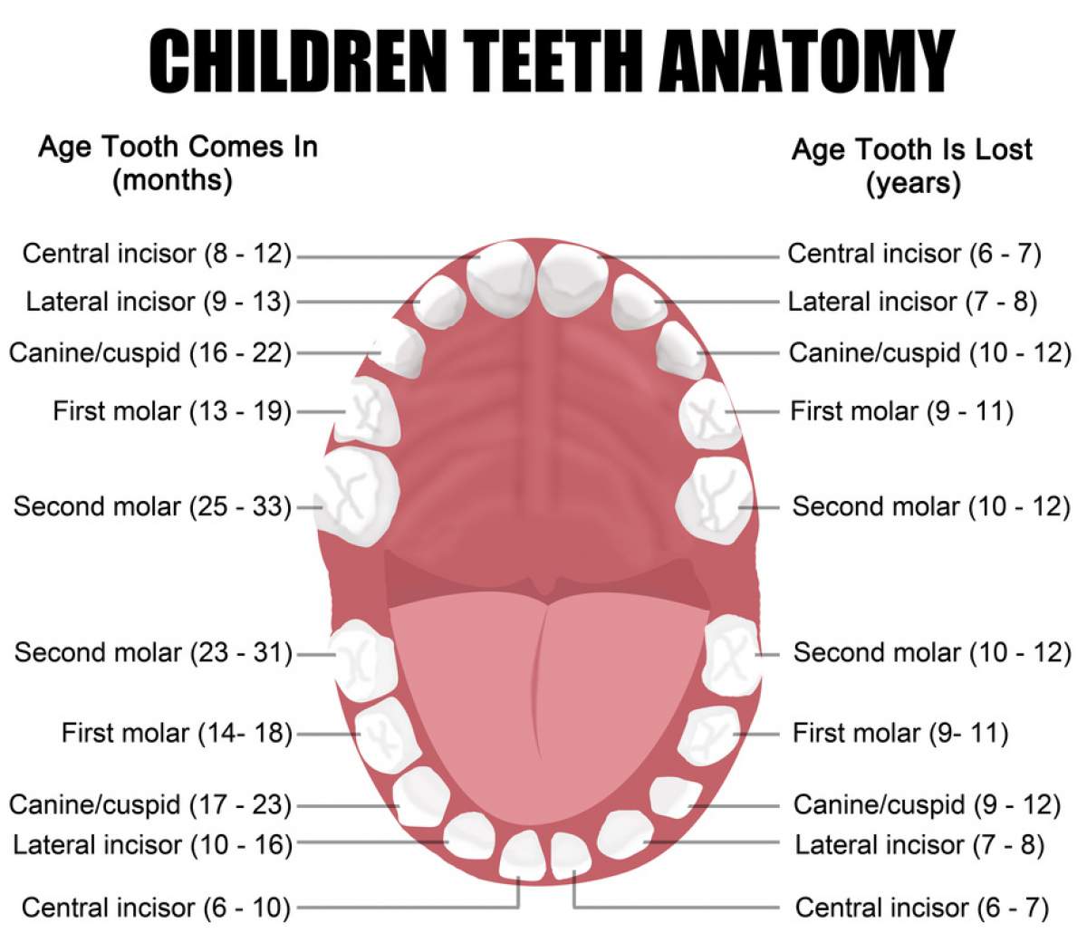
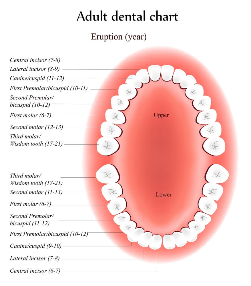
A View of the Mouth Inside the Tooth 3D MODEL Tooth eruption There is a broad range of normal times for teeth to push through the gum tissue (erupt) into the mouth. For primary teeth, the central incisors are the first teeth to erupt, occurring at about 6 months of age.

Mouth Teeth Diagram with Label Health Images Reference
The mandible and maxilla - like most bones in the human body - have a core of less dense cancellous bone, wrapped in an outer layer of more dense alveolar bone. The part of the mandible and maxilla that are in the mouth are covered by the gums. And the teeth rest in bony sockets within the mandible and maxilla and are surrounded by the gums.
Children Teeth Anatomy Dentist Manhattan NY
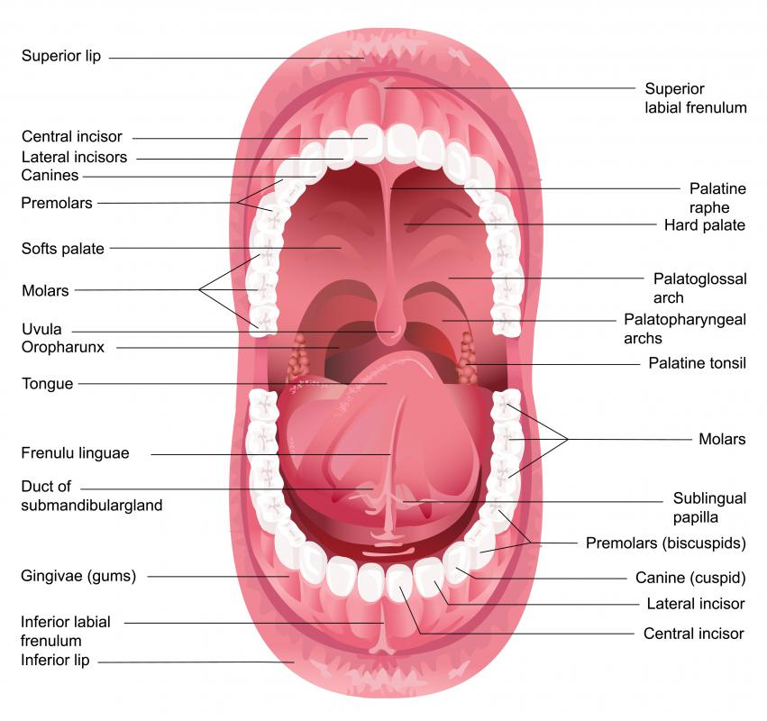
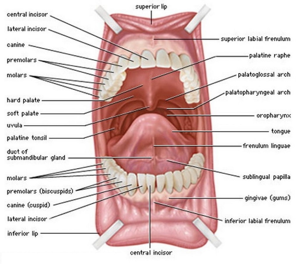
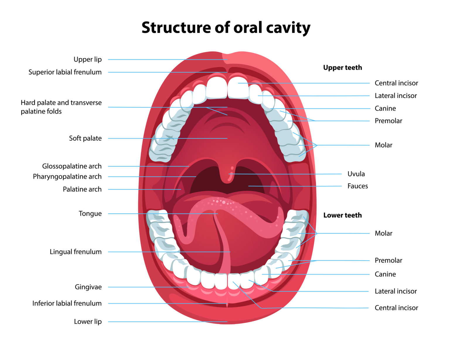
The oral cavity, or more commonly known as the mouth or buccal cavity, serves as the first portion of the digestive system. It consists of several different anatomically different aspects that work together effectively and efficiently to perform several functions. These aspects include the lips, tongue, palate, and teeth. Although a small compartment, the oral cavity is a unique and complex.
Mouth Diagrams Printable 101 Diagrams
Place a pea-sized dab of fluoride toothpaste on the head of the toothbrush. (Use a soft toothbrush.) Place the toothbrush against the teeth at a 45-degree angle up to the gum line. Move the brush.
What Are the 3 Key Functions of the Teeth? Vancouver Centre for
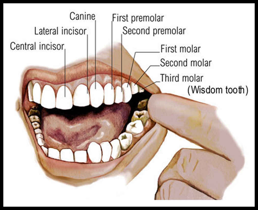
Learn about the types of teeth in a fast and efficient way using our interactive tooth identification quizzes and labeled diagrams. This leaves up to eight adult teeth in each quadrant and separates the opposing pairs within the same alveolar bone as well as their counterparts in the opposing jaw. Each quadrant contains: a medial incisor
The Purpose of Teeth Dr. Kevin Sands
Roof The roof of the mouth proper consists of the hard and soft palates. The hard palate is found anteriorly. It is a bony plate that separates the nasal cavity from the oral cavity. It is covered superiorly by respiratory mucosa (ciliated pseudostratified columnar epithelium) and inferiorly by oral mucosa (stratified squamous epithelium).

Dental Mouth Diagram for Education Health Images Reference
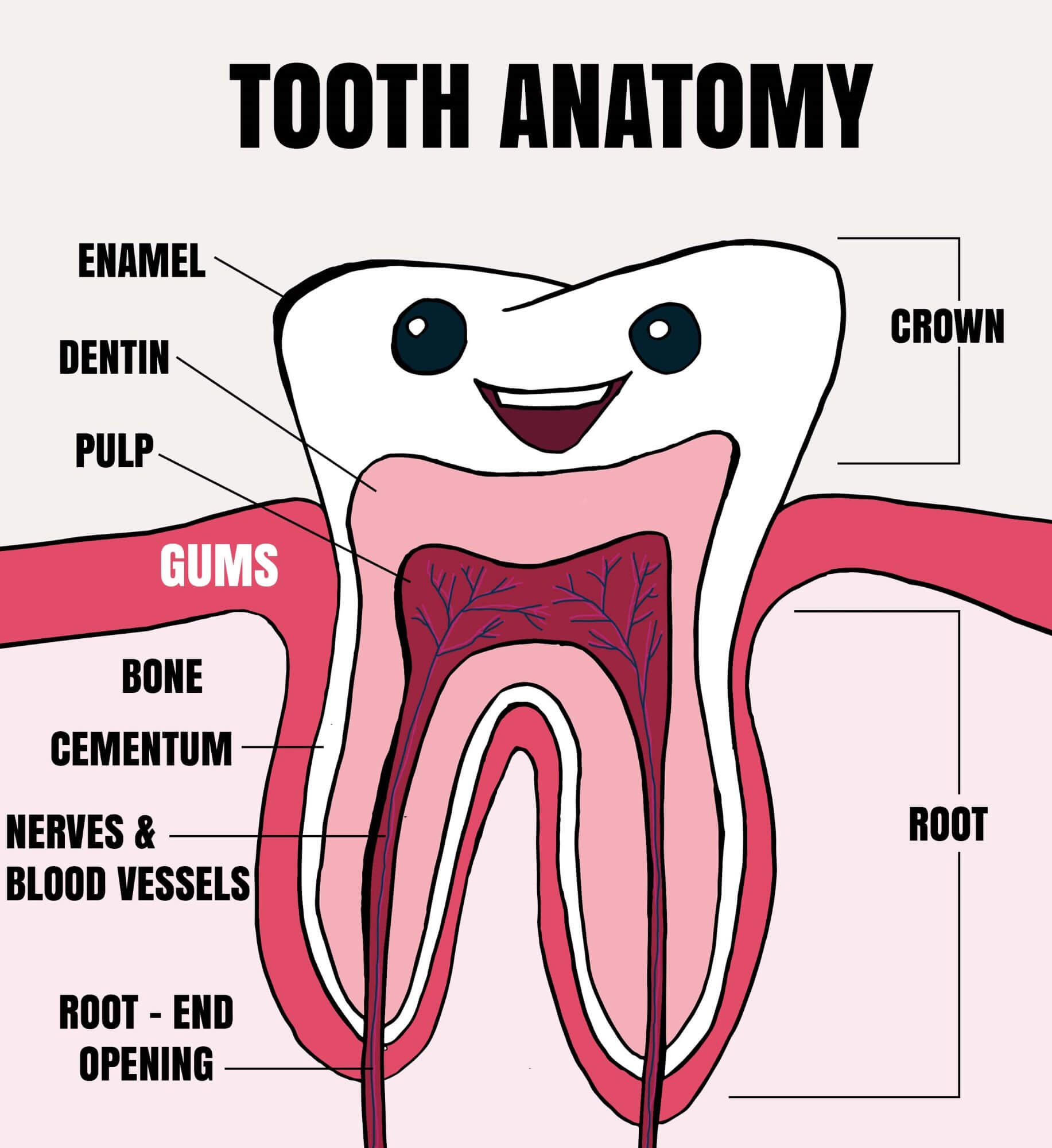
It's made up of several parts: Root canal. The root canal is a passageway that contains pulp. Cementum. Also called cement, this bone-like material covers the tooth's root. It's connected to the.
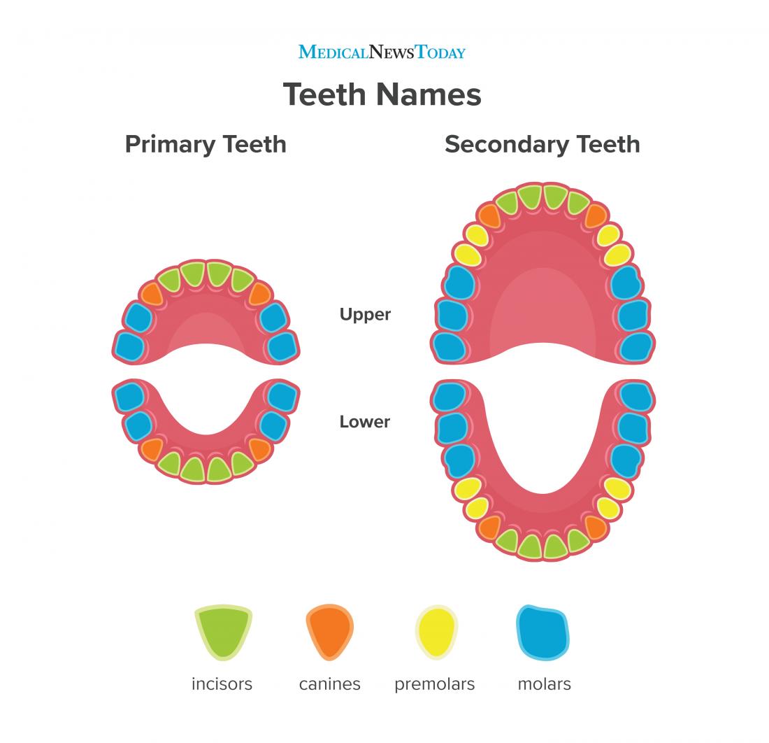
Teeth names Diagram, types, and functions
What's my mouth's function? Your mouth supports many daily functions, including: Breathing. Talking. Chewing. Tasting. Swallowing. Eating. Drinking. Mouth function in digestive system Your mouth is where digestion begins. When you chew food, your salivary glands make saliva (spit). Saliva helps break down starches in the foods you eat.

Mouth Teeth Diagram with Label Health Images Reference
Figure 1. Teeth numbers and names diagram. The human teeth is composed of 16 upper teeth and 16 lower teeth. They are also divided into four quadrants. Have you ever struggled reading your dental treatment plan from your local dentist? Sometimes, it feels like deciphering a difficult table from a college statistics book.
Mouth Teeth Diagram with Label coordstudenti
To record changes to your dental health, dentists use a chart with a diagram of your teeth. The teeth are numbered according to the Universal Numbering System adopted by the American Dental Association. The diagram is drawn as if you're looking at your dentist with your mouth wide open. The top teeth are numbered from right to left.
Tooth Anatomy Explained for Kids Tooth Fairy Smiles Blog
In this section, you will examine the anatomy and functions of the three main organs of the upper alimentary canal—the mouth, pharynx, and esophagus—as well as three associated accessory organs—the tongue, salivary glands, and teeth. The Mouth. The cheeks, tongue, and palate frame the mouth, which is also called the oral cavity (or buccal.

Mouth Teeth Name In Human Health Images Reference
Adult teeth are called permanent or secondary teeth: 8 incisors. 4 canines, also called cuspids. 8 premolars, also called bicuspids. 12 molars, including 4 wisdom teeth. Children have just 20.
Dental Articles and Resources
Canines Canines are the sharp, pointed teeth that sit next to the incisors and look like fangs. Dentists also call them cuspids or eyeteeth. Canines are the longest of all the teeth, and people.

23.3 The Mouth, Pharynx, and Esophagus Anatomy & Physiology
Enamel. The outer layer of the tooth and the hardest material in the body. Dentin. The inner layer and the main part of the tooth, and the largest dental tissue. Pulp. Soft tissue on the inside of the tooth that contains the nerve, blood supply, and the ability to produce dentin. Root. The part of the tooth that secures it into the jaw.